 Fraunhofer Institute for Cell Therapy and Immunology IZI
Species
Mouse
Fields of application
In the xenogeneic murine tumor model cancer cell lines or primary patient cells are injected into immunodeficient Nude mice so that they form a tumor. Human as well as murine cells can be used for this purpose. The tumor can be precisely irradiated after CBCT scan or the scan is used alone for noninvasive imaging. By using cell lines expressing luciferase tumor growth can be noninvasively visualized and quantified by bioluminescence imaging (BLI). Thus the model provides the opportunity to test the potential of novel anti-cancer therapeutics.
- Pharmacodynamics and pharmacokinetics
- (Patho)physiological processes
- Therapeutic efficacy
- Proof of concept
Endpoints / outcome parameter
- Clinical symptoms (tumor growth; in vivo)
- Hemogram analysis (in vivo)
- Tumor (patho)histology (ex vivo)
Readout parameter
- Cone beam computed tomography (CBCT)
- Measurement of tumor size (by means of caliper)
- Bioluminescence imaging (BLI)
- Magnetic resonance imaging (MRI)
- Animal blood counter
- Flow cytometric analysis
- RT-PCR
- Western Blot
- Histology (various classical histological stains)
- Immunohistochemistry
Quality management and validation
- Controls
- Blinded induction
- Blinded data collection and analysis
- Randomisation
- Allocation concealment
- Biometric expertise
- Internal quality management
References
Commissional work for Tavarlin AG (Darmstadt, Germany). "Inhibition von TKTL1 in einem murinen Tumormodell."
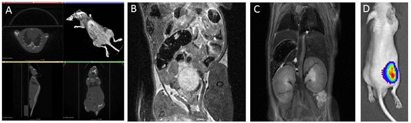
Different noninvasive imaging options for tumors and other applications available at the Fraunhofer IZI. A: CBCT scan of a mouse. The 2D projections are used to generate a 3D Image. B+C: MRI of a mouse B shows the abdominal area, in image C the lung and kidneys are visualized. D: Visualization of a tumor by means of bioluminescence imaging (BLI). Cells are injected into the flank of the mice. Already 7 days later a distinct luminescent signal can be detected.