Microscopy

© Fraunhofer IZI

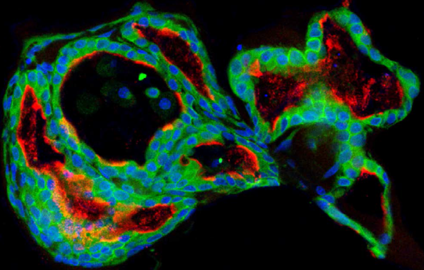
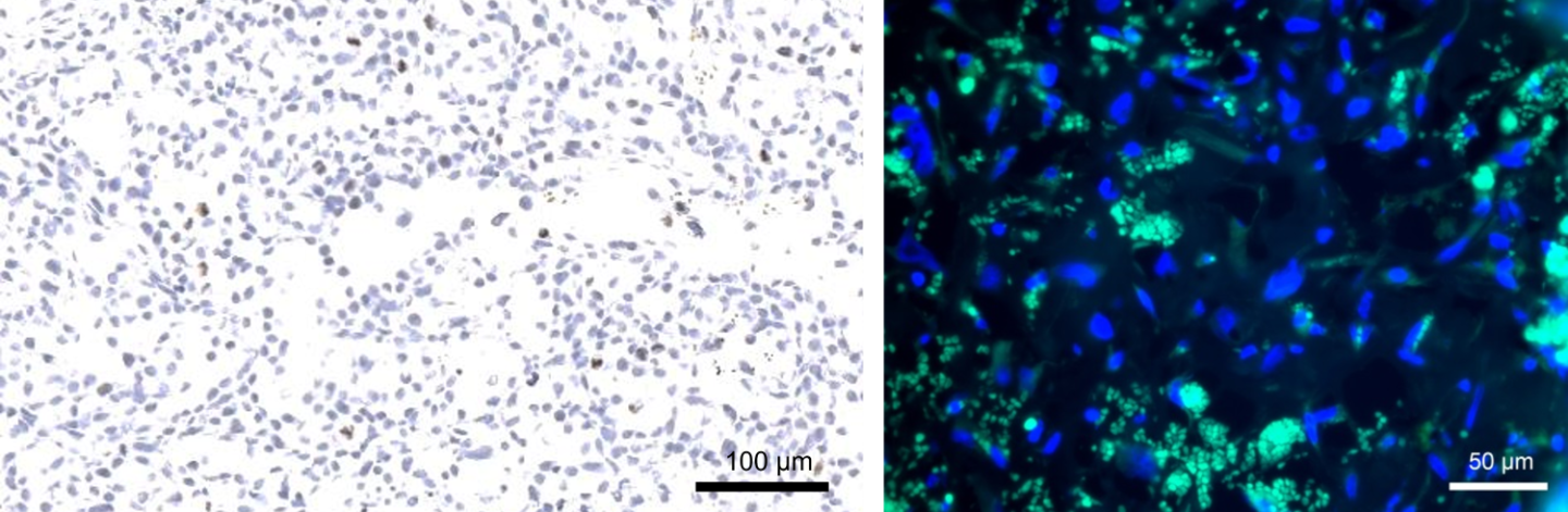
Phenotyping biological samples using multiple imaging methods forms a core competence of preclinical research. This enables thorough depiction, from the smallest structures (cell organelles) right through to entire organ systems, both in spatial and temporal resolution (4D). Fraunhofer IZI has access to a comprehensive, state-of-the-art equipment pool that enables the acquisition and evaluation of various (also correlative) image data. Partners and customers are advised on biological, technical and economic matters and supported in carrying out and evaluating experiments. Furthermore, experimental procedures and equipment can be used, adapted and developed.

The following methods and devices are offered. Detailed device overviews can be found on the pages of the Experimental Imaging Unit.

Methods and devices

Brightfield, fluorescence and automated slide scanning microscopy

  • Research Microscope Nikon Eclipse Ti
    • All standard contrast methods (brightfield, phase contrast, fluorescence) and magnifications (5x-100x)
  • Slide Scanner Zeiss Axio Scan.Z1
    • Automatic scanning of slides
    • Online access to the digital scans
    • Sending of the scans on a data carrier
    • Slide scanning under GLP conditions

Live cell microscopy

  • Microscope Zeiss Axio Observer.Z1
    • Automated atmospheric conditions (temperature, humidity and CO2 content)
    • 6D microscopy (2D images, z-focus, time series, positions and image stiching)
  • Live cell analysis platform Sartorius IncuCyte® SX5
    • Data acquisition directly in the cell culture incubator
    • Inserts for cell culture flasks, multi well plates, petri dishes and slides
    • Cell samples remain stationary and only the optics for data acquisition move. Allows gentle observation of sensitive samples
    • Automatic evaluation of a wide range of assays (e.g. cell growth, viability, cytotoxicity, ...)

Confocal Laser Scanning Microscopy

  • Microscope Zeiss Axio Oberserver.Z1 with LSM 710
    • Laser excitation at 405, 458, 488, 514, 532 and 633 nm
    • Automated atmosphere conditions (see live cell imaging above)
    • 6D microscopy (see live cell imaging above)

In-vivo imaging

  • Magnetic resonance imaging (MRI) in the Bruker Pharmascan 7T for small animal
  • Computed tomography (CT) and radiation planning in the Xstrahl SARRP for small animals
  • Bioluminescence (BLI) and fluorescence imaging (FLI) in the IVIS Spectrum and Spectral Instruments Imaging Ami HT for small animals
  • Imaging of large animals with partners of the University of Leipzig