Sepsis in huNSG mice

Species

mouse

Fields of application

Sepsis is a life-threatening disease but even today it lacks targeted drugs that improve the therapy of affected patients. To study reactions of human immune cells in vivo we use NSG mice transplanted with human hematopoietic stem cells (HSC) to engraft a functional human immune system. After maturation of the human immune system in these mice they become injected with lipopolysaccharide (LPS) and develop clinical symptoms of sepsis.

  • Pharmacodynamics and pharmacokinetics
  • (Patho)physiological processes
  • Therapeutic efficacy
  • Proof of concept

Endpoints / outcome parameter

  • Score (severity of clinical symptoms; in vivo)
  • Immune cells in full blood (in vivo)
  • Cytokines, antibodies and protein levels (all human and murine) in blood plasma (in vivo)
  • Cellular infiltrates in and pathophysiology of divers organs (ex vivo)

Readout parameter

  • Scoring
  • Flow cytometry
  • ELISA / CBA (cytometric bead array)
  • RT-PCR
  • Western Blot
  • Histology (various classical histological stains)
  • Immunohistochemistry

Quality management and validation

  • Controls
  • Blinded induction
  • Blinded data collection and analysis
  • Randomisation
  • Allocation concealment
  • Biometric Expertise
  • Internal quality management

References

Scholbach J, Schulz A, Westphal F, Egger D, Wege AK, Patties I, Köberle M, Sack U, Lange F. Comparison of hematopoietic stem cells derived from fresh and cryopreserved whole cord blood in the generation of humanized mice. PLoS One. 2012; 7(10):e46772.

Rodewohl A, Scholbach J, Leichsenring A, Koeberle M, Lange F. Humanized NSG mice with immature immune cells react qualitatively different on LPS stimulation than mice with mature human cells. Innate Immun. 2016; Submitted.
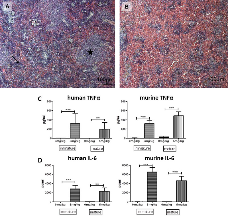
Symptoms of sepsis in humanized NSG mice. A+B: Histological sections of spleens stained with H&E (Hematoxylin and Eosin stain). A shows a spleen of a representative mouse treated with saline. Regularly developed white pulp with lymphocytic follicles is visible (asterisk) sinusoids are covered by lymphocytes (arrow) and few erythrocytes. B: Spleen of an animal treated with 5 mg/kg LPS. The mouse was euthanized 6 hours after treatment. White pulp is extremely diminished while red pulp dominates. Lymphocytic follicles are decreased in size and erythrocytes fill the space between sinusoids. C+D: The levels of both human and murine TNFα and IL-6 in blood plasma were analyzed by cytometric bead array. All cytokines are strongly increased 6 hours after injection of LPS compared to control animals injected with saline. ** p ≤ 0.01; *** p ≤ 0.001.
© Fraunhofer IZI
Symptoms of sepsis in humanized NSG mice. A+B: Histological sections of spleens stained with H&E (Hematoxylin and Eosin stain). A shows a spleen of a representative mouse treated with saline. Regularly developed white pulp with lymphocytic follicles is visible (asterisk) sinusoids are covered by lymphocytes (arrow) and few erythrocytes. B: Spleen of an animal treated with 5 mg/kg LPS. The mouse was euthanized 6 hours after treatment. White pulp is extremely diminished while red pulp dominates. Lymphocytic follicles are decreased in size and erythrocytes fill the space between sinusoids. C+D: The levels of both human and murine TNFα and IL-6 in blood plasma were analyzed by cytometric bead array. All cytokines are strongly increased 6 hours after injection of LPS compared to control animals injected with saline. ** p ≤ 0.01; *** p ≤ 0.001.