NSG mice with xenogeneic immune system

Species

Mouse

Fields of application

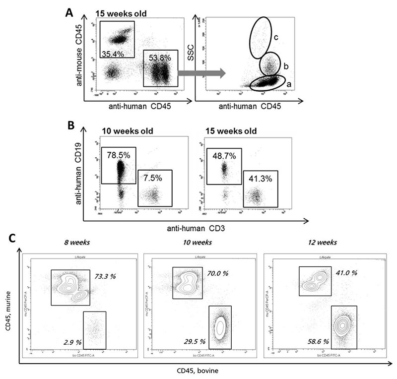
For the engraftment of xenogeneic immune systems the NSG mouse is currently the gold standard host because of its high grade immunodeficiency. In our group hematopoietic stem cells from different species and sources (e.g. cord blood, bone marrow) are used for this purpose. Newborn mice become preconditioned and afterwards transplanted with these cells. Within some weeks after transplantation, all main immune cell subpopulations develop in the NSG mice and constitute a functional xenogeneic immune system. After 8 to 10 weeks the xenogeneic, differentiated immune cells can be detected and quantified by flow cytometric analysis.

This model perfectly fits the demands of infection studies, the study of autoimmune and tumor diseases, and inflammatory research for the graft species.

  • Pharmacodynamics and pharmacokinetics
  • (Patho)physiological processes
  • Therapeutic efficacy
  • Proof of concept

Endpoints / outcome parameter

  • Immune cells in full blood (in vivo)
  • Cytokines, antibodies and protein levels (all murine and graft species) in blood plasma (in vivo)
  • Histology of divers organs (ex vivo)

Readout parameter

  • Flow cytometry
  • ELISA / CBA (cytometric bead array)
  • RT-PCR
  • Western Blot
  • Histology (various classical histological stains)
  • Immunohistochemistry

Quality management and validation

  • Controls
  • Blinded induction
  • Blinded data collection and analysis
  • Randomisation
  • Allocation concealment
  • Biometric Expertise
  • Internal quality management

References

Scholbach J, Schulz A, Westphal F, Egger D, Wege AK, Patties I, Köberle M, Sack U, Lange F. Comparison of hematopoietic stem cells derived from fresh and cryopreserved whole cord blood in the generation of humanized mice. PLoS One. 2012; 7(10):e46772.

Development of xenogeneic immune cells in NSG mice. A: Newborn mice are transplanted with human hematopoietic stem cells. Peripheral blood is analyzed by means of flow cytometric analysis at different time points after transplantation. The left dot plot shows leukocytes of a successfully transplanted mouse at 15 weeks after transplantation. Human and mouse leukocytes can be differentiated. Within all living leukocytes, 35.4 % are murine and 53.8 % have human origin. In the right dot plot human CD45+ cells are plotted against sideward scatter (SSC). Population (a) represents lymphocytes whereas population (b) are monocytes and population (c) represents granulocytes. B: Percentages of huCD45+ subpopulations differ during time course after transplantation. Dot plots depict representative percentages of huCD19+ and huCD3+ within population of huCD45+. Ten weeks old animals show higher levels of huCD19+ compared to 15 weeks old animals but a lower amount of huCD3+ in comparison to the older mice. C: Flow cytometric analysis of peripheral blood in the time course after transplantation of a newborn NSG mouse with bovine hematopoietic stem cells. Percentages of bovine CD45+ cells substantially increase with time after transplantation.
© Fraunhofer IZI
Development of xenogeneic immune cells in NSG mice. A: Newborn mice are transplanted with human hematopoietic stem cells. Peripheral blood is analyzed by means of flow cytometric analysis at different time points after transplantation. The left dot plot shows leukocytes of a successfully transplanted mouse at 15 weeks after transplantation. Human and mouse leukocytes can be differentiated. Within all living leukocytes, 35.4 % are murine and 53.8 % have human origin. In the right dot plot human CD45+ cells are plotted against sideward scatter (SSC). Population (a) represents lymphocytes whereas population (b) are monocytes and population (c) represents granulocytes. B: Percentages of huCD45+ subpopulations differ during time course after transplantation. Dot plots depict representative percentages of huCD19+ and huCD3+ within population of huCD45+. Ten weeks old animals show higher levels of huCD19+ compared to 15 weeks old animals but a lower amount of huCD3+ in comparison to the older mice. C: Flow cytometric analysis of peripheral blood in the time course after transplantation of a newborn NSG mouse with bovine hematopoietic stem cells. Percentages of bovine CD45+ cells substantially increase with time after transplantation.