Specialist group Image Analysis of Cell Function successfully evaluated
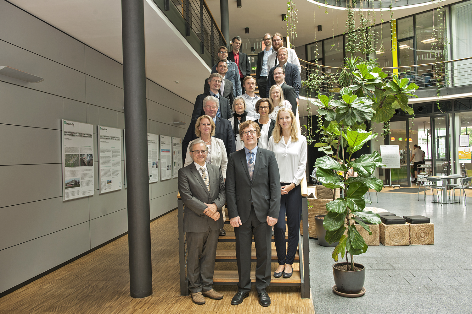
On 12 August 2014, the Fraunhofer Institute for Cell Therapy and Immunology and Leipzig University of Applied Sciences (HTWK Leipzig) got the ball rolling on a joint specialist group focused on the “Image Analysis of Cell Function”. The group was evaluated and its future viability assessed on 4 August 2017, following a three-year start-up phase. The evaluation committee, made up of representatives from science, business and politics, attested to the outstanding work completed by the team, which is headed up by Professor Ulf-Dietrich Braumann, during this early phase and unanimously recommended that the specialist group be continued. The group's high level of commitment to the teaching and training of young scientists also received a special mention.


The specialist group was put together as part of a cooperation program with the universities. The program aims to help universities strengthen their profile and expand training opportunities besides giving students access to a high-quality research infrastructure and taking a hands-on approach to familiarizing them with application-oriented research. For the cooperating Fraunhofer institutes, expanding core research areas, broadening the range of business services and appealing to up-and-coming scientists all lie at the heart of the initiative. In order to set up the Leipzig specialist group, the Fraunhofer-Gesellschaft provided 1.5 million euros in funding for a five-year period. HTWK Leipzig introduced the “Biotronic Systems” chair for the duration of the project, supported by the Saxon State Ministry for Science and the Arts.
Professor Ulf-Dietrich Braumann brought both the chair and the concept of the joint specialist group to life. From the very start of the cooperation, students from HTWK Leipzig were involved in the specialist group located at Fraunhofer IZI and were acquainted with competitive research on an international scale. Furthermore, six scientific members of staff from Fraunhofer IZI contributed towards teaching activities at HTWK Leipzig on the topics of bioreactors and microscopic imaging. Through these activities, more and more electrical engineering and information technology students began taking an interest in life science matters while engineering skills were introduced and utilized at Fraunhofer IZI. This led to four projects researching practical approaches and also five master’s theses being completed within the group during the start-up phase alone. An additional five master’s theses and a doctoral research project are currently under way.
The scientific focus of the specialist group lies on establishing and further developing a non-destructive imaging technology for biomedical research. Before new therapeutic approaches can be created, new drugs and their effect on living and damaged tissue first have to be validated in order to demonstrate healing and protective effects. These effects are, however, very difficult to follow over periods of several hours and even days using image-guided procedures. The light intensity and distribution of conventional microscopy procedures damage the study samples and lead to cell death in the case of long-term observations. In addition, existing markings (fluorochrome) are bleached out, resulting in distorted findings. The working group therefore set itself the task of establishing a system at the institute for the non-destructive long-term monitoring of living organisms, tissue and cells. Improvements and new developments in the field of light sheet microscopy are central here.
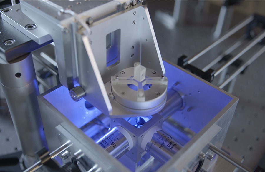
Well-known since 2004, light sheet fluorescence microscopy (LSFM), also referred to as selective plane illumination microscopy (SPIM) or light sheet microscopy, provides an imaging method which is much more gentle on samples, also over long periods of time from hours and days through to weeks. The main advantage of this technique is the special light geometry which facilitates a better distribution of light intensity across the sample. The light stress placed on the individual cells is thus significantly reduced, as is the bleaching of labeling dyes. The advantages of light sheet microscopy in terms of conducting long-term studies and the enormous variability of the system were the deciding factors in setting up the institute’s own light sheet microscope at Fraunhofer IZI. Special attention was paid here to the types of image data that are able to be generated. Three-dimensional image data records can be created through the systematic movement (translation, rotation) of a biological sample, allowing depth information to be acquired from samples that has so far not been possible using confocal microscopy.
Besides the SPIM technology, the specialist group will also work within the research and business units “Biomedical Image Analysis”, “Biosignal Analysis” and “Industrial Image Analysis” in future. The goals for the coming year are to entrench the group within the Fraunhofer model, to significantly boost staff and to expand teaching and research activities.
Contact
Prof. Dr. Ulf-Dietrich Braumann
Head of Image Analysis of Cell Function Unit
Phone +49 341 35536-5416
ulf-dietrich.braumann@izi-extern.fraunhofer.de