Contact Press / Media
Dr. Natalia Sandetskaya
Research scientist MicroDiagnostics Unit
Fraunhofer Institute for Cell Therapy and Immunology
Perlickstr. 1
04103 Leipzig, Germany
Phone +49 341 355369310
Fax +49 341 355369930
The aim of this project is to evaluate the economic prospects for innovative applications of regionally produced special materials in in vitro diagnostics. Fiber-based and / or porous materials, which were originally used primarily in the manufacture of textiles or building materials, could have many advantageous properties for sample preparation in laboratory diagnostics. Their implementation for new purposes would provide an ideal opportunity for innovation in diagnostics and materials manufacturing, while also opening up a new market for materials manufacturers in Saxony.
Around 75 % of all cervical cancer cases are caused by the human papilloma virus. Vaccines are available to prevent this; however, these are not used comprehensively. In addition, the vaccination is only effective provided there was no prior persistent infection. This means a fast, effective and reliable test could help healthcare systems to stratify the persons to be vaccinated and, on the other hand, it could increase awareness of the need for screenings which facilitate the earlier detection of infections and cancers and can improve the success rate in treatment.
This project aims to develop a point-of-care test which takes up sample material and releases the nucleic acids of the viral pathogens. These are replicated via isothermal amplification with the required temperature being achieved and maintained with the help of an exothermic chemical reaction. Afterwards, the result of the detection reaction can simply be read off. Moreover, the cartridge is to be made of a sustainable material which, in turn, is manufactured from renewable raw materials and is compostable.
Fraunhofer IZI contributes its expertise in sample processing and the development of molecular biological rapid tests.
Through its gynecology clinic, Leipzig University Hospital will become involved in the project as a clinical partner on the basis of a subcontract. In addition to medical expertise, it will also contribute clinical samples of patients with an HPV infection in order to evaluate the functionality of the system in a proof-of-concept study.
Project partners
INTU Diagnostics GmbH (coordination); VOXDALE GmbH; Leipzig University Hospital Leipzig AöR; Institut für Molekulare Diagnostik und Bioanalytik (IMDB) gGmbH
Point-of-care diagnostics (such as Covid rapid tests) are usually designed for broad application outside specialized medical facilities and produced in large quantities. The majority of the materials used for this are single-use, oil-based plastics. The enormous raw material consumption, lack of recycling and the materials used (which are poorly biodegradable) are causing enormous environmental problems.
Bio-based and bio-degradable plastics are obviously a much more sustainable alternative. Novel bio-based plastics (such as PLA, HNA, TPS) are usually more difficult to process and, for this reason, they are seldom used in the diagnostics industry at present.
The project aims to develop a suitable, bio-based and, if possible, bio-degradable material as well as to establish corresponding process chains and manufacturing processes for the new materials. The general suitability and functionality of this material is examined using the example of a hepatitis D point-of-care rapid assay.
Fraunhofer IZI contributes its expertise in the development and validation of molecular biological and immunological tests as well as microfluidic systems. During the project, the institute is responsible for the development of a lateral flow assay for the fast and reliable detection of virus proteins and antibodies in the serum or plasma. To enable this, regulatory aspects for the approval of medical products are already being considered during the development phase.
Project partners
University od Applied Sciences Dresden (coordination); Otto Injection Molding GmbH & Co. KG; Roboscreen GmbH; Bergi-Plast GmbH; Fraunhofer Institute for Machine Tools and Forming Technology IWU
The aim of the project is to develop an easy-to-use test system for the on-site analysis (point-of-care) of clinically relevant biomarkers in blood. Similar to a blood glucose measurement, the analysis should be fully automated and based on just a few drops of blood.
The detection approach is based on a class of bioluminescent synthetic sensor proteins developed by the Max Planck Institute for Medical Research, which change the color of the emitted light in the presence of the analyte. This light can be captured by a powerful camera, as is already used in cell phones today, and quantified via the ratio of the emitted light colors.
One of the core tasks of the Fraunhofer IZI within the project was the development of a microfluidics-based test platform with automated sample processing. This should be designed in such a way that only a few manual pipetting or dilution steps are necessary, making the process user-friendly even for non-experts. In addition to the biosensors, the necessary reagents must also be provided in a chemically stable form within the test system by means of freeze-drying. The sample is transported exclusively by capillary forces, which minimizes the use of electronics and mechanics. This reduces manufacturing costs and increases mobility and system stability.
To produce the microfluidic cartridges, three different methods were initially evaluated: 3D printing, flexographic printing and hot embossing, with the latter proving to be the most suitable method.
An assay for analyzing nicotinamide adenine dinucleotide (NAD+) was then implemented on this microfluidic test platform. NAD+ plays an essential role in various biochemical processes. A decrease in NAD+ levels has been associated with various pathologies and physiological ageing, while strategies to increase cellular NAD+ levels have been effective against age-related diseases in many animal models.
In the final phase of the project, the assay will be validated using patient samples.
The project is funded by the cooperation program between institutes of the Max Planck Society and the Fraunhofer Gesellschaft.
Project partner
Max Planck Institute for Medical Research; Fraunhofer Institute for Applied Optics and Precision Engineering IOF
Within the last years the interest in extracellular vesicles (EVs) increased dramatically due to their capability to represent their cell of origin during the moment of release. Within our group we investigate this feature as a promising approach for diagnostics in liquid biopsy. Although EVs are only 30 nm up to a few micrometer in size they are enriched in multiple molecules (proteins, nucleic acids, lipids, metabolites, among others). Those potential biomarkers are surrounded by a lipid bilayer protecting them against extracellular degradation. In addition, EVs are continuously secreted from virtually all cells and are consequently found in all bodily fluids (e.g. blood plasma, urine, saliva). Thus, non- to minimally-invasive isolation is possible and conventional tissue biopsy could be avoided in the future.
At the Fraunhofer IZI an antibody microarray was established to analyze the expression of EV surface markers in malignant diseases from different bodily fluids. The findings are valuable for the enrichment of tissue- or disease specific EVs. The aim is to separate the relatively small fraction of disease-associated EVs from the broad background of "healthy" EVs and to study their cargo. A correlation within the EVs and the tumour cells is expected, however, additional changes in the profile might be identified which could improve or perhaps even render diagnostic or therapeutic decision making possible in the future.
The aim of the project is to research molecular acoustics applications for the quantitative and qualitative classification of blood, blood plasma and dialysis filtrates. The project will also look at how emitted acoustic waves propagate in bodily fluids. Specific characteristics will be identified that pinpoint the tiniest differences in the composition of the investigated medium. Moreover, it will also be characterized to what extent multisensor systems used in vivo are able to carry out non-destructive real-time analyses and therefore surpass existing diagnostic procedures in terms of speed and precision. Besides acoustic sensors, optical and magnetic sensors are also to be used.
This project is co-financed by tax revenues on the basis of the budget approved by members of the Saxon state parliament.
The diagnosis of diseases should be as quick, easy and inexpensive as possible, directly at the point of care without highly qualified laboratory personnel and also without additional stress for the patient. To meet the existing requirements, research is being carried out at the Fraunhofer IZI and the Fraunhofer Center MEOS on a diagnostic method using exhaled air.
Breathing gas analysis has been used clinically for years as part of lung function tests or the urea breath test to detect Helicobacter pylori infections. However, there is a much greater potential in breathing gas diagnostics. There are thousands of so-called volatile organic compounds (VOCs) whose composition in the air we breathe changes with certain diseases and infections. For example, the diabetic patients’ smell of overripe fruit is known, caused by a greatly increased concentration of acetone, a VOC.
Most VOCs occur in extremely low concentrations (ppmV to pptV - one particle per million or trillion particles). This places extreme demands on the measurement technology. Conventionally, mass spectroscopic methods are mostly used, which, however, mean an enormously high expenditure on equipment. We therefore focus on ion mobility spectrometry (IMS), which is coupled with gas chromatographic (GC) pre-separation. This technology is much cheaper, more portable (shoebox size) and is already being used for drug and explosives controls.
In Leipzig, GC-IMS was able to differentiate between different bacteria based on their VOC profile after only 90 minutes of cultivation in the laboratory in 2019. In the course of the project, these experiences will now be used to directly examine real breath samples in the clinic. The aim is to diagnose various bacterial and viral infectious diseases, including any existing drug resistance, in patients’ breath.
In the Fraunhofer Center MEOS in Erfurt, colleagues from Fraunhofer IZI and Fraunhofer IPMS are working on a new IMS system. The centerpiece is a small IMS silicon chip, which is being further developed at Fraunhofer IPMS in Dresden. Potentially miniaturized IMS chips can be inexpensively manufactured in large quantities using the microelectronic manufacturing processes. The new system is to be tested in the diagnosis of neurological diseases and cancer. In addition to the exhaled air, other non-invasive samples such as sweat and urine are also in focus.
Explanation of the IMS measurement procedure which is being developed at the Fraunhofer Center MEOS. Video realization © 3D Agentur Berlin, Dirk Puder und Stefan Loth, GbR
Exhaled air contains substances known as volatile organic compounds (VOCs), which provide information about metabolism. In a variety of diseases, including infections, cancer and neurodegenerative diseases, the metabolism and thus the composition of the exhaled VOCs changes. Detection of these VOCs offers the opportunity to diagnose diseases early and non-invasively.
Ion mobility spectrometry (IMS) can detect VOCs within minutes directly at the point-of-care. The BMBF project ”Breath Alert” investigates whether IMS can be used to detect antibiotic resistance in bacteria. In the Fraunhofer-versus-Corona cluster project ”M3Infekt”, IMS technology was further developed at the Fraunhofer Center for Microelectronic and Optical Systems for Biomedicine (MEOS) with the participation of Fraunhofer IZI. Specifically, methods for sampling via mouth and nose, for short-term sample preservation and for sample preparation were established and tested. At the end of the project, the method was tested on 60 healthy volunteers in two clinical studies in Dresden and Magdeburg. In parallel, a functional novel IMS demonstrator was completed at MEOS. This must now be further developed and optimized in follow-up projects to selectively detect diagnostics-relevant VOCs in complex matrices such as exhaled breath.
In the M3Infekt project, the participating nine Fraunhofer Institutes developed further non-invasive and mobile sensors for recording heart rate, ECG, oxygen saturation, respiratory rate and respiratory volume. Concepts for system integration and flexible interfaces were defined and a multimodal AI framework for cross-sensor data evaluation was developed. In addition, requirements regarding conformity to medical regulatory requirements were developed. The overall vision of the project is a close monitoring of relevant clinical parameters for detecting condition deterioration in infectious diseases also outside of intensive care units via a multimodal, modular and mobile sensor system. During the project it has become apparent that several specific solutions for different sub-applications are more useful than a single overall system and thus the benefit of the project results is even increased.
In a report, the World Health Organization emphatically describes why the fight against antibiotic resistance is one of the major tasks faced by the global community. By the year 2050, the organization expects 10 million deaths annually that will be attributable to infectious agents [1]. Innovations are necessary not only for therapeutic treatment, but also for the diagnostic detection of pathogens that cause disease to counteract the challenge in the healthcare sector.
The BreathAlert project launched at the end of 2020 aims at improving this situation with a new method for the rapid and non-invasive detection of infectious agents and antibiotic resistance, which analytically analyzes patients’ breath. The project focuses on the further development of ion mobility spectrometry, which is to be used to characterize volatile organic components (VOCs) of microorganisms.
At the Fraunhofer IZI, selected microorganisms are being examined to determine whether they can be differentiated via the VOCs released and whether they can be assigned to the respective types of bacteria. For this purpose, the pathogens are first cultivated and then the headspace, the gas phase above the culture medium, is fed into the device. The VOCs are ionized, separated in an electric field and then detected at different times. Software analyzes the complex data. The aim is to identify specific signals with which bacteria can be reliably differentiated from one another, even under different conditions. The focus is on antibiotic-resistant pathogens, such as enterobacteria, which are increasingly showing resistance to carbapenem and cephalosporin [1].
The characterization of clinical isolates, air samples from infected patients and measurements of the influence of e.g. eating habits on the air we breathe round off the project.
The company Graupner medical solutions GmbH, which develops the medical device technology, works together with the Fraunhofer IZI as a consortium partner. The development work is supported by specialist clinics that provide access to samples and also carry out a final validation. The project results are used economically by the Graupner company.
1] World Health Organization Report 2017: Prioritization of Pathogens to guide discovery, research and development of new antibiotics for drug-resistant bacterial infections, including tuberculosis. WHO/EMP/IAU/2017.12
![BMBF_CMYK_Gef_M [Konvertiert]](/en/departments/leipzig-location/infection-research-diagnostics/microdiagnostics/projects/jcr:content/contentPar/sectioncomponent_442397282/sectionParsys/imagerow_379030831/imageComponent1/image.img.jpg/1626434958831/BMBF-gefoerdert-2017-en.jpg)
Sample preparation is a crucial aspect in many areas of bioanalytical research, especially in the analysis of the crude complex samples and/or rare targets. Modern laboratories exploit very sensitive methods of detection including molecular diagnostics; however, their performance strongly depends on the quality of the sample. Pre-analytical processing must prepare the specimen for the most effective detection of the target. It includes the purification of the analyte, its pre-concentration, as well as the parallel removal of the compounds which may affect the analysis. The preservation of samples which prevents the degradation of the target is also a task for this field of applied analytics. The main aim of sample preparation is to ensure the precision of the subsequent analysis. None of the sample preparation approaches are universal: they must take into account methods and equipment for the downstream processing of the specimen, concentration and nature of the analyte, volume of the sample and many other factors.
Nowadays bioanalytical research actively pursues integrating complex assays on automated platforms including lab-on-chip devices. This trend often lacks intelligent solutions for the pre-analytical steps. The aim of our working group is to support this particular field by developing of the most suitable sample preparation approaches for specific needs.
The group supports researchers and industrial partners with customized solutions, evaluates capacities of existing methods and develops novel strategies for effective pre-analytical processing.
The significance of easy-to-use test strip systems for the rapid detection of clinically relevant parameters or for quality assurance of food products is increasing not only in developing countries. We develop a simple diagnostic platform that is particularly suitable for nucleic acid-based formats. As a reference assay, pathogens are diagnosed in human samples.
Modern air traffic is not just useful for the fast transport of passengers or goods. Even infectious agents travel by planes over long distances within a few hours as unwanted passengers. Infectious diseases such as influenza or SARS, which can develop into pandemics, are nowadays spreading rapidly and much faster than years ago. Steps to effectively control and prevent chains of infection in the field of modern mobility have not yet been effectively established worldwide. Initial approaches using simple questionnaires or non-contact temperature measurement, however, have remained largely ineffective. The project "HyFly", funded by the German Federal Ministry of Education and Research (BMBF) within the InfectControl 2020 initiative, addresses the sensitive issue of passenger control. Together with partners, a non-invasive method based on ion mobility spectrometry (IMS) is being worked on. Applying this strategy, infected persons should be identified within a few minutes via components of their breathing air. IMS is already routinely used worldwide to detect drugs or explosive remnants in airports providing an established infrastructure. So-called volatile organic substances that are metabolic products of microorganisms are detected. The focus is on bacterial pathogens, which, according to information from the German Robert Koch Institute, have a high relevance for aviation. Initial results show that the method has great potential for discriminating between different pathogens. In addition to system development, a study is underway to identify microorganisms at various international airports to determine the cleaning efficiency and impact of antimicrobial coatings.
Periodontitis is an inflammatory disease of the gums that, if left untreated, can lead to tooth loss. In Germany alone it is predicted that nearly 12 million people are affected by periodontitis. The main trigger for periodontal disease is bacterial plaque which can lead to a reduction of the dental bone tissue. The postulated systematic relationship between periodontal disease caused by bacterial pathogens and cardiovascular damage has been studied extensively. It can result in particularly serious diseases such as heart attacks and strokes.
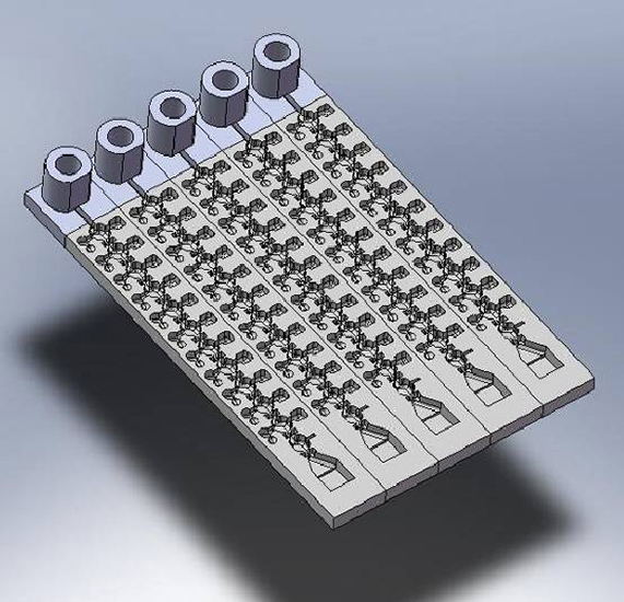
The parodontitis chip project is aimed at developing a fully integrated diagnostics platform both for the fast processing and the subsequent analysis of periodontal pathogens in complex samples. This innovative technology consists of a compact microfluidic card and a combined purification module. Steps such as isolating pathogenic nucleic acids, selectively amplifying DNA sequences, and their specific detection are integrated to establish an easy-to-use setup for the end-user.
The lab-on-a-chip device will allow the detection and characterization of 11 bacteria relevant to the pathogenesis of periodontitis in a parallel format. In addition, the establishment of a simple detection unit will allow the monitoring of reaction kinetics. Therefore a quantification of the pathogen, as well as a determination of the total bacterial count can be realized.
The parodontitis chip project will allow for the creation of a simple molecular diagnostic test platform that can easily be adapted to various problems in the field of medical, environmental, or food analysis. Simplified lab-on-chip devices having an extremely simple structure and non-contact detection units provide significant time and cost savings for the user.
Tuberculosis is an infectious disease, which is caused by Mycobacterium tuberculosis. According to a 2013 report of WHO (World Health Organisation), Tuberculosis ranks as the second leading cause of death from an infectious disease worldwide, after the human immunodeficiency virus (HIV).
An early and reliable diagnosis is crucial. Our unit is currently developing in cooperation with the McMaster University in Hamilton (Canada) a detection system which is rapid, simple, and cost-efficient. The nucleic acid-based test system integrates all steps from pathogen isolation to hybridization of nucleic acid on pathogen-specific probes.
Athletes have been trying to improve their performance through illegal means for decades. They use specific products that can enhance endurance, muscle growth and strength or improve recovery after extensive trainings/contests.
Within this project, a diagnostic device is developed to detect different doping products in blood samples. A complete integration of sample preparation and bioassay will be designed and developed. Furthermore, the power of this device will be the detection of multiple doping products in one assay based on surface plasmon resonance (SPR) technology.


A reliable diagnosis of complex and life-threatening infectious diseases (e.g. sepsis) is currently only possible using elaborate and time-consuming methods involving an analysis laboratory and qualified specialists. The Unit is developing an innovative system for rapid, easy-to-conduct and inexpensive on-site infection diagnostics.
The system is based on magnetic particles in the micrometer scale that can be functionalized according to their respective application to act as carriers for antibodies and disease-associated DNA sequences. These magnetic particles are employed on a disposable object in the approximate shape of a check card. In an on-site examination a sample is obtained from the patient, such as blood, saliva or urine, which is then incorporated into the lab-on-a-chip system. After lysis of the target cells, the magnetic particles bind to the respective target molecules in the sample and are transported via magnetic forces through different reaction tubes in a fully automated manner. At the end of the process chain the diagnosis is performed using a highly sensitive magnet sensor system.
The project is funded by the Federal Ministry for Education and Research (BMBF, Bundesministerium für Bildung und Forschung) and coordinated by Magna Diagnostics GmbH, a spin-off company of the Fraunhofer IZI.
Circulating tumor cells (CTCs) are cancer cells shed from a primary tumor and circulated in the peripheral blood. Detection and isolation of CTCs can be used for diagnostics purposes and e.g. personalized medicines. Circulation tumor cells (CTCs) differ from peripheral blood mononuclear cells (PBMCs) in many ways. Detecting CTCs in blood samples is however a challenge, since CTCs comprise a very rare population among red blood cells and leukocytes.
The ApoStream™ technology from ApoCell can detect and enrich CTCs from blood with the use of a method called dielectrophoresis (DEP). This device is still in development and in order to validate the system, surrogates for CTCs and PBMCs need to be developed. Both biological as non-biological surrogates are designed, produced, screened and optimized.
Reporter gene assays offer a wide range of applicabilities in modern research. In the first place, they serve for characterizing regulatory elements (promoter regions) or modulators (transcription factors) on the genomic level. In particular, luciferase-based reporter gene assays are established in current biomedical and pharmacological research. Due to their extremely low detection limit they have prevailed over fluorescence-based reporter genes. Basically, the regulatory genetic element to be investigated is cloned upstream of a luciferase gene. The reporter gene construct can be introduced into a selected cell system by means of conventional transfection methods. The biological activity of the cloned genetic element can now be characterized in a time-dependent manner. The Fraunhofer IZI offers a complete dual-luciferase system that is suitable for the investigation of genetic regulatory elements from mammalian genomes.
The method comprises three steps:
optional steps:
In a cooperation project with the department of General Biochemistry of the University of Leipzig we succeeded in investigating the activity of the human GLO1 promoter in different carcinoma cell lines. At present, the influence of hypoxia on promoter activity is being investigated.
The system is used for the functional characterization of any conceivable regulatory element from mammalian genomes. Moreover, the system offers a way of examining exogenous factors in combination with normal or altered culture conditions.
RNAi is a conserved mechanism that regulates gene expression on the post-transcriptional level. In eukaryotes, double-stranded RNA (dsRNA) is processed to form short, small interfering RNAs (siRNA) which results in the degradation of the complementary mRNA. Finally, this leads to the effective down-regulation of the specific protein. These characteristic features result in the application of RNAi in cell cultures and animal models. The specific suppression of gene expression and protein activity modulates the pharmacological inhibition of the target protein and is thus an effective tool in proof-of-principle experiments and the identification and validation of antitumor agents.
For providing a novel cancer therapy, the reference project aims at the specific and gradual down-regulation of an already identified target protein known as glyoxalase I (GLO I) by means of RNAi. Moreover, the system shall provide a way of re-up-regulating GLO I in order to recreate the initial phenotype.
In tumors there is a strong correlation between signal transduction pathways and fundamental metabolic pathways, such as glycolysis and the pentose phosphate cycle. Many tumor-promoting mechanisms have an immediate effect on glycolysis, on the cellular response to oxygen and on the ability of tumors to recruit new vessels for their own nourishment. Since Warburg it has been known that tumors consistently utilize anaerobic pathways to produce ATP by conversion of glucose. One cytotoxic byproduct of glycolysis is methylglyoxal (MGO). The reactive MGO binds to proteins and nucleic acids in high concentrations, thereby inducing apoptosis. All organisms have a glyoxalase enzyme system (GLO I / GLO II) to prevent cell damage caused by high MGO concentrations. In particular in tumor cells, this system is up-regulated in order to minimize the paracatalytically generated high MGO concentrations. The inhibition of glyoxalases could therefore play an important role in cancer therapy. To date, the characterization of the glyoxalases in malignant tumors only referred to histochemical analyses which show an overexpression in tumors. By means of the RNAi model, specific GLO I inhibitors could be identified in substance libraries.