 Fraunhofer Institute for Cell Therapy and Immunology IZI
We provide a cell-based model of the human gut epithelial barrier to analyze the impact of test substances and/or infectious agents on gut barrier integrity and function. Caco-2 cells (human epithelial cell line) and THP-1 cells (human monocytic cell line) are co-cultured in a transwell culture system that models the epithelial barrier (Caco-2 cells) and the underlying lamina propria with resident macrophages (THP-1 cells). After addition of test substances and/or infectious agents to the upper chamber of the transwell system ("gut lumen"), epithelial barrier integrity and function as well as transmigration of pathogens and the resulting immune response by epithelial cells and macrophages is analyzed using various approaches.
In vitro model for gut epithelial barrier (GLP-like / GLP)
Species
Human
Fields of application
- Preclinical drug development
- Intestinal infection studies
- Toxicity studies
- Efficacy and safety studies for drug approval (GLP-like and GLP)
Endpoints / outcome parameters
- Barrier integrity / permeability
- Cell viability / cytotoxicity
- Pathogen invasiveness / migration
- Profile of cytokines / chemokines
- Changes in tissue marker expression (e.g. tight junction proteins)
- Ultra-structural analysis of epithelial barrier
Readout parameters
- TEER (Transepithelial electrical resistance)
- FITC-Dextran test
- Cell viability / cytotoxicity assays
- CFU assays
- ELISA / Multiplex analysis
- Histology
- Immunohistochemistry / immunofluorescence
- Transmission electron microscopy
Quality management and validation
- Internal quality management (certified GLP test facility)
- Use of reference compounds
- Randomisation
- Blinded induction / data acquisition and analysis
- Biometric expertise
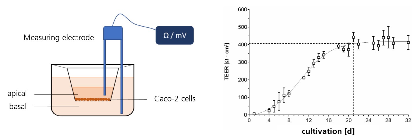
TEER measurement of Caco-2 cells in a transwell system.
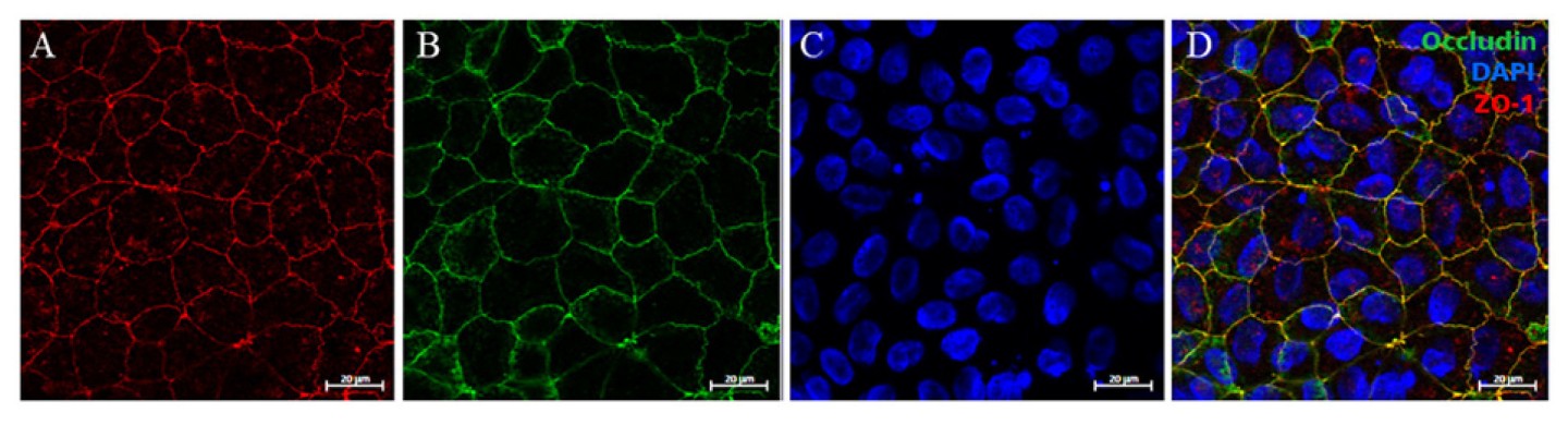
Indirect immunofluorescence staining of tight junction proteins from untreated Caco-2 cells; (A) staining of membrane proteins ZO-1 (red), (B) occludin (green), (C) control staining of cell nuclei with DAPI (blue), (D) representation of total staining; scale bar: 20 μm; magnification 63x.