Hitchhiker’s Guide to the Cell: Fraunhofer IZI researches to improve imaging methods for organoids
On 1st March 2020, a new research project was launched at the Fraunhofer Institute for Cell Therapy and Immunology (IZI). Imaging of living cells is to be improved by means of viral and bacterial targeting sequences. The project is supported by the VolkswagenStiftung.

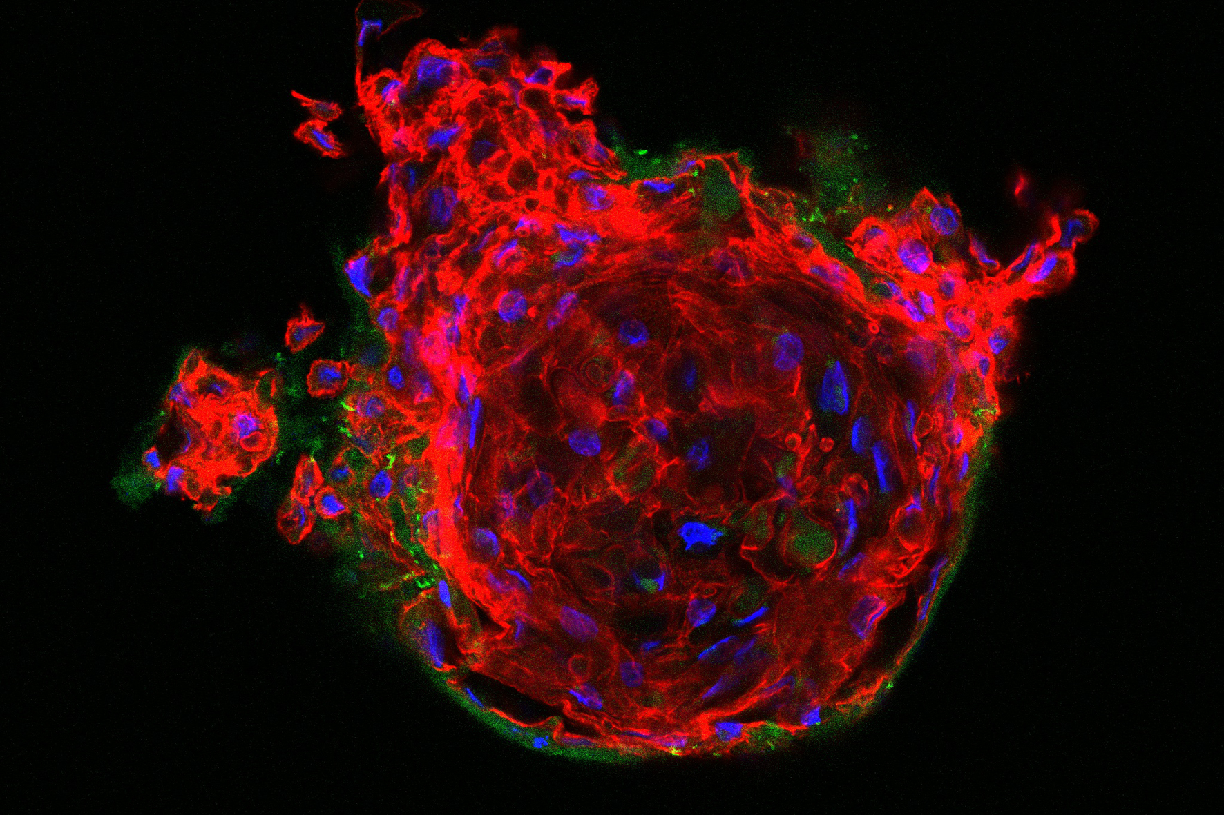
Organoids are three-dimensional cell cultures. These precursors of organs can be produced in test tubes and permit the examination of vital processes in the human organism outside the body. As a result, they are considered to be an optimum solution for closing the gap between 2D lab cultures of human cells and animal tests.
“In recent years, the development of organoids has made rapid progress. However, at the moment, analysis tools for the continuous examination of the processes in organoids are still missing”, explains Dr Claire Fabian, a stem cell biologist at Fraunhofer IZI. Dr Fabian runs the project together with Dr Sebastian Geiser, a physicist and head of the Fraunhofer IZI working group on experimental imaging. In the framework of the “Hitchhiker’s guide to the cell” project, methods are to be established which, as it were, permit “hitchhiking” through human cells. Greiser, who contributes his expertise on imaging of three-dimensional structures, adds: “We want to develop methods in order to visualise the spatial and temporal dynamics of complex 3D cell cultures. To this end, we will fine-tune the life-science and technology questions throughout the entire project.”
The project team is planning to analyse the specific interactions of viral and bacterial proteins and peptides with cellular components. The scientists hope to use the insights obtained to develop imaging instruments. In this context, they are focussing, in particular, on organelles. These are specific compartments within a cell that fulfil a certain function, e.g. mitochondria which produce energy. If the scientists are successful with their concept, which is based on the idea of dyeing specific organelles in the living cells, it will be possible to visualise cellular processes of novel and complex 3D cell culture systems and then use these to “hitchhike” through the cells. Ultimately, the aim is to improve our understanding of the mechanisms, on which, e.g., development biology, modelling of illnesses and regenerative research are based.

Over a period of one and a half years, the project “Hitchhiker’s guide to the cell – Employing viral and bacterial targeting sequences for live cell imaging” will receive funding of roughly EUR 120,000 from the VolkswagenStiftung in the framework of the programme “Experiment! – Auf der Suche nach gewagten Forschungsideen” [Experiment! – Looking for ground-breaking research ideas] supporting daring and innovative research ideas that fundamentally challenge existing established ways of thinking.

