Fraunhofer Institute for Cell Therapy and Immunology IZI
Species
Mouse
Fields of application
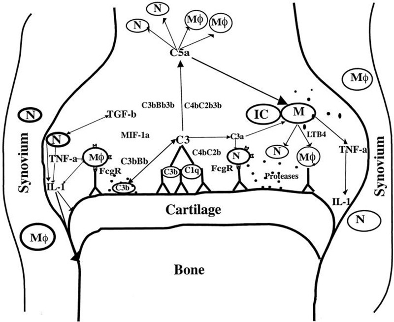
Collagen antibody induced arthritis (CAIA) in mice is a well characterized animal model for rheumatoid arthritis (RA) in man. CAIA is induced by intravenous injection of antibodies directed against epitopes of collagen II, followed by a booster injection with an adjuvant. CAIA is an acute model for RA. However, the adaptive immune response is only playing a minor role in this model. The whole inflammatory process is mediated by complement and the innate immune system. About 14 days after onset clinical symptoms decline. Assessment of clinical symptoms is realized by means of an established scoring system.
- Pharmacodynamics and pharmacokinetics
- (Patho)physiological processes
- Therapeutic efficacy
- Proof of concept
Endpoints / outcome parameter
- Score (severity of symptoms; in vivo)
- Immune cells in full blood (in vivo)
- Cytokines and RA marker protein levels in blood plasma (in vivo)
- Swelling, destruction, pannus formation, immune cell infiltration (ex vivo)
- Halogenating activity of myeloperoxidase (MPO; in vivo)
Readout parameter
- Scoring
- Flow cytometry
- ELISA / CBA (cytometric bead array)
- RT-PCR
- Western Blot
- Histology (various classical histological stains)
- Immunohistochemistry
- MPO activity assay
Quality management and validation
- Controls
- Blinded induction
- Blinded data collection and analysis
- Randomisation
- Allocation concealment
- Biometric Expertise
- Internal quality management
References
Lange F, Bajtner E, Rintisch C, Nandakumar KS, Sack U, Holmdahl R. Methotrexate ameliorates T cell dependent autoimmune arthritis and encephalomyelitis but not antibody induced or fibroblast induced arthritis. Ann Rheum Dis. 2005; 64(4):599-605.
Flemmig J, Schwarz P, Bäcker I, Leichsenring A, Lange F, Arnhold J. Rapid and reliable determination of the halogenating peroxidase activity in blood samples. J Immunol Methods. 2014; 415:46-56